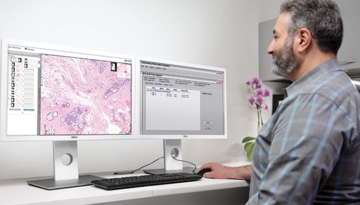
Confidence in Diagnosis for the Pathologist
The Aperio GT 450 DX starts with quality and stays with quality. High quality digital scans deserve an image management system that detects, stores and delivers whole slide images and metadata exactly where and when needed for confidence in diagnosis.
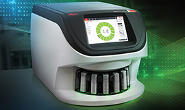
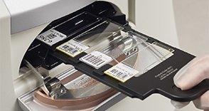
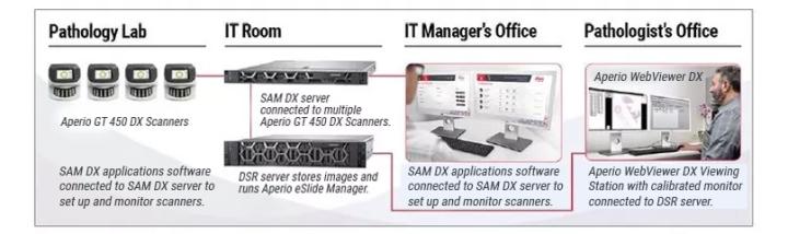
Aperio GT 450 DX Scanner
- Designed to rapidly deliver images with exceptional quality for Pathologists.*
- High performance objective manufactured by Leica Microsystems, renowned for producing world-class optics since 1849.
- Leica optics deliver exceptional image quality
- Every slide is calibrated during scanning to ensure the best quality image
- Automated image quality assessment, so you don’t have to send slides back for re-scans 99.5% accurate tissue finder (H&E stains) finds faint tissue while excluding pen marks, dust, and residue.


")


")