
Aperio GT 450 DX

Aperio GT 450 DX scanner, WebViewer DX, eSlide Manager and LIS Connectivity

Proven technology for improved workflow efficiency and patient care are set to define digital pathology and increase overall adoption.

The Aperio GT 450 DX is a comprehensive digital pathology system. It partners the time-tested Aperio GT 450 DX scanner together with a secure and integrated image management system - complete with a clinically validated clinical viewer and LIS/LIMS connectivity. State-of-the-art svs and DICOM imaging, excellent image quality, and robust LIS connectivity capabilities allow for a seamless and secure integration with hospital systems for a streamlined and efficient workflow.

With interoperability at the forefront for this solution, the Aperio GT 450 DX scanner is an in vitro diagnostic device for use with compatible viewing software and display that has been validated as an aid to the pathologist in the qualitative review and interpretation of digital images. Aperio WebViewer DX is a viewing software application for use with digital images from compatible Aperio GT 450 DX scanners on local and remote networks.

Contact us to get a quote. Call +1 844 534 2262 to speak with a representative.

For In Vitro Diagnostic Use.

Confidence in Diagnosis for the Pathologist

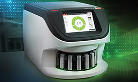
The Aperio GT 450 DX starts with quality and stays with quality. High quality digital scans deserve an image management system that detects, stores and delivers whole slide images and metadata exactly where and when needed for confidence in diagnosis.

Aperio GT 450 DX Scanner

  • Designed to rapidly deliver images with exceptional quality for Pathologists.** 
  • High performance objective manufactured by Leica Microsystems, renowned for producing world-class optics since 1849.
  • Leica optics deliver exceptional image quality
  • Every slide is calibrated during scanning to ensure the best quality image
  • Automated image quality assessment, so you don’t have to send slides back for re-scans
  • 99.5% accurate tissue finder (H&E stains) finds faint tissue while excluding pen marks, dust, and residue.

Aperio WevViewer DX with eSlide Manager and LIS Connectivity

  • Compare current case with past patient cases to provide needed context, with the ability to synchronize image magnification and rotation of multiple images within the same window
  • Corroborate your diagnosis with colleagues and consulting sub-specialists using advanced communication features
  • Streamline slide navigation for professional quality presentations using handy Pin Drops
  • LIS Integration
  • Image Enhancement
  • Macro View to detect missing tissue
  • Multi-level sort for slide tray images
  • Measurements in microns/mm

**See Resources > DICOM for more info on How it Works with the Aperio GT 450 DX

Image
Confidence in Diagnosis for the Pathologist

Security and Interoperability for IT Professionals

The Aperio GT 450 DX system offers fast, secure, flexible IT architecture

Aperio GT 450 DX Scanner:

  • HL7, svs and DICOM compatible for easy integration into LIS, LIMS or PACS
  • A centralised SAM DX server and dedicated software provides you with a data management solution that can remotely set up and monitor multiple Aperio GT 450 DX scanners at a time.
  • Comprehensive cybersecurity including encryption and login controls
  • Centralised scanner management to set up and monitor multiple scanners
  • Scalable set up for hub and spoke configurations
  • Supports dedicated logins for each Aperio GT 450 DX scanner

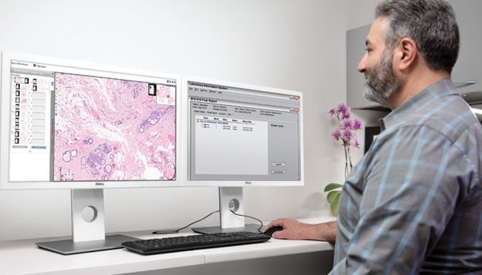
Aperio WebViewer DX with eSlide Manager and LIS Connectivity

  • Microservices-based architecture
  • Hub and spoke network architecture
  • Supports MS Server Windows 2016 and 2019
  • Single gateway for users across multiple locations
  • Enhanced cybersecurity: PHP v7, updated Apache, OpenSSL for Data encryption between browser and system web pages, and complex passwords
  • In-app user authentication and authorization ensure that the right people have access to the right functionality and data
  • Browser-based thin client
  • Supports SVS Images
  • Integrate your system to allow secure access to existing hospital infrastructure
Image
Security and Interoperability for IT Professionals

Speed to Quality for Lab Managers and Histotechnicians

The Aperio GT 450 DX is simple to operate and helps lab managers and histotechnicians deliver rapid, quality results with confidence

Aperio GT 450 DX scanner:

  • No touch continuous loading during scanning
  • Automated IQ checks during each scan to ensure quality
  • Assign priority cases
  • Each slide is calibrated individually prior to scan
  • 450-slide capacity packed into a small size fits on the lab bench
  • 99.5% accurate tissue finder (H&E Stains) allows you to adjust if needed*
  • 100% first-time barcode scan success rate*
  • Slides are then automatically scanned at 81 slides per hour at 40x for a 15mm x 15mm area.
  • Fast delivery of urgent cases directly to the pathologist’s viewing station

*VP-0463 validation results

Image
Speed to Quality for Lab Managers and Histotechnicians The Aperio Clinical Solution is simple to operate and helps lab managers and

Resources

 
Aperio GT 450 DX interactive demo

Frequently Asked Questions

Aperio GT 450 DX scanner
How many slides per hour can it scan?

81 slides per hour @ 40x (15mm x 15mm area).

What is the scan speed?

32 seconds at 40x for 15mm x 15mm area

Does it have a workstation?

No, Aperio GT 450 DX has a scalable IT architecture solution …where a centralized SAM DX server is used to set up and monitor multiple GT 450 DX scanners from the IT professionals' office.

What is a SAM DX server?

Scanner Administration Manager DX (SAM DX) is a high-performance server installed in the IT room that enables a centralized admin hub that is connected to multiple GT 450 DX scanners in the pathology lab. The GT 450 DX scanners stream raw image files to the SAM DX server after which they are converted to an SVS file. SAM DX includes Mirth for data and event handling and HL7 for integration. SAM DX server client application software is loaded on the SAM DX server and enables IT professionals and lab directors to log in via secure network access and then set up and monitor GT 450 DX scanners in the lab, including dashboard status views, PIN controls for each scanner and log out times for each scanner.

Can I adjust the tissue finder?

Yes, in the rare case you need to adjust the tissue finder, the user can simply select “Scan Entire Slide” and the Aperio GT 450 DX will automatically scan the entire slide.

Do I need to perform pre-snaps?

No, this is automated with the Aperio GT 450 DX because it uses Real-Time Focusing.

Do I need to use calibrated monitor?

Yes, Aperio GT 450 DX images are designed to be viewed on calibrated monitors for the best possible viewing experience as it relates especially to color. The Aperio WebViewer DX, includes monitors that are calibrated and have been validated for use with images generated by Aperio GT 450 DX scanners. The Aperio WebViewer DX includes a proprietary image color correction (ICC) profile that calibrates the monitor for use with Aperio GT 450 DX images.

Monitor Manufacturer Monitor Model Number Supported Settings
Dell UP3017 Factory settings
Dell U3223QE Brightness set to 90
Barco N.V. MDPC-8127 Factory settings.
Dell U3023E Factory settings.
Is every slide calibrated during scanning?

Yes, during each scan, the scanning workflow includes a system calibration. This calibration is included in the 32 sec scan speed at 40x for a 15mm X 15mm area.

What is the resolution?

0.26 µm/pixel

Does it offer continuous loading while scanning?

Yes. This unique open face design allows up to 14 additional racks to be added or removed while scanning is in progress.

Are the Aperio GT 450 DX slide racks compatible with other devices from Leica Biosystems

Yes, these racks can be used across 3 of our devices, including Aperio GT 450 DX, HistoCore SPECTRA workstation ST Stainer, and HistoCore SPECTRA workstation CV Coverslipper.

DICOM
What is DICOM file format?

DICOM (Digital Imaging and Communications in Medicine) is a standard for medical image interchange that includes support for transmission and storage of medical image data.

How is DICOM used?

It can be used to transfer data from a Picture Archiving and Communication System (PACS) into some other system, produced by any number of vendors who choose to implement that standard, or used to provide access to clinical data without granting that person access to the entire PACS, or to share medical image data with a patient or colleague, or for long-term archival purposes.

Why is DICOM important?

DICOM is an open, vendor-agnostic standard for medical image data. While many vendors participate in the creation of this standard, it is not controlled by any one vendor. As an open standard, it may be implemented by any vendor and enables interoperability between systems.

Is there a Universal DICOM file for Digital Pathology?

The DICOM standard provides a definition for Whole Slide Microscopy Images, commonly referred to as a “Supplement 145 image” (after the DICOM Supplement which initially defined the image) or “Working Group 26 image” (after the DICOM Working Group which produced Supplement 145). As this standard is intended to be usable by manufacturers of both tiling and line scanning systems, as well as different modalities (e.g. oil, fluorescence, brightfield), it provides vendors multiple ways to produce images that are conformant to the standard, but may not necessarily be compatible with a given vendor’s PACS or viewing software.

Downloads

Case Studies & White Papers

Aperio LIS Connectivity English