![Leica VT1000 S Vibrating blade microtome Leica VT1000 S Vibrating blade microtome](/sites/default/files/styles/thumbnail_fallback/public/media_image-file/2021-03/Leica-VT1000-S-Vibrating-blade-microtome.jpg?itok=mWTw-hnd)
![Adjustable 2x large-field magnifier Adjustable 2x large-field magnifier](/sites/default/files/styles/thumbnail_fallback/public/media_image-file/2021-03/VT1000S_Magnifier_knife_holder_R_43.jpg?itok=1lLdEL9w)
![Magnetic specimen plate Magnetic specimen plate](/sites/default/files/styles/thumbnail_fallback/public/media_image-file/2021-03/csm_vt1000S_magnetic_specimen_clamp_43_704d369c62.jpg?itok=N729uGCw)
![Transversal section through the rat brain cortex at the forebrain level. A large single neuron was labeled by NADPH-diaphorase histochemistry. The small axon and some branching dendrites are visible. (Source: Dr. Andreas Schober, University of Heidelberg Transversal section through the rat brain cortex at the forebrain level. A large single neuron was labeled by NADPH-diaphorase histochemistry. The small axon and some branching dendrites are visible. (Source: Dr. Andreas Schober, University of Heidelberg](/sites/default/files/styles/thumbnail_fallback/public/media_image-file/2021-03/csm_VT9B_46_5b252c57c6.jpg?itok=tVJF3-Ky)
![CA 3 field of rat hippocampus CA 3 field of rat hippocampus. Syn. positive axon terminals over pyramidal cells.](/sites/default/files/styles/thumbnail_fallback/public/media_image-file/2021-03/CALB16_46.jpg?itok=p9zyBP86)
![Rat Brain Neurophysin-immunoreactice (ir) neurons shown in a transversal section through the paraventricular nucleus of the rat brain. (Source: Dr. Andreas Schober, University of Heidelberg, Dept. of Neuroanatomy & IZN)](/sites/default/files/styles/thumbnail_fallback/public/media_image-file/2021-03/VT1B_46.jpg?itok=nWj-c6f3)
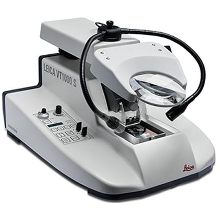
라이카 VT1000 S 진동 블레이드 박절기
Leica VT1000 S 진동 블레이드 박절기는 신경생리학, 신경병리학, 실험병리학,식물학(뿌리 및 초목) 및 산업(폼) 분야의 고품질 절편 제작 요구 충족을 위해 선택하는 장비입니다.
특정 신경병리학 시험을 위해고정된 조직의 절편을 제작할 때 Leica VT1000 S는 고품질 절편 결과물을 일관되게 제공합니다.
사용자 문의
견적이 필요하시면
연구용으로만 사용하십시오. 진단 절차에는 사용하지 마십시오.
기술 사양
일반사항
최대 검체 크기: | 70 x 40 x 15 mm |
블레이드 이동 속도: | 0.025 - 2.5 mm/s |
조절 가능한 절삭 윈도우: | 전자식 |
총 수직 시료 스트로크: | 15 mm |
확대 옵션: | 확대경 2개 |
진폭: | 0.2; 0.4; 0.6; 0.8; 1.0 mm |
총 수직 시료 스트로크: | 15 mm(전동식) 표준 |
절편 제작 범위: | 1~40mm(조절 가능) |
제품의 기능
주파수와 진폭을 변경할 수 있습니다.
다양한 애플리케이션 요구 사항을 신속하게 수용하기 위해 사용자는 0~100Hz 사이에서 주파수를 변경하고 0.2~1mm 사이에서 0.2mm 단위로 적절한 진폭을 선택할 수 있습니다.
조정 가능한 나이프 진행 속도
이 기기는 0.025 ~ 2.5mm/s 사이에서 매우 정밀하게 조정 가능한 나이프 진행 속도, 프로그래밍 가능한 절편 창 및 표본 후퇴 기능을 갖추고 있습니다.
완전 자동 절단 모드
절편 프로세스 가속화: Leica VT1000 S는 고속 나이프 리턴 스트로크와 자유롭게 프로그래밍할 수 있는 절편 창을 갖추고 있어 가장 작은 시편도 매우 빠르게 절편할 수 있습니다.
고품질 절편
조정 가능한 시편 후퇴 기능은 나이프의 리턴 스트로크 중 시편 손상을 최소화합니다.
다운로드
Brochure
Certificate
IFU and SDS
구성
기본형 기기 (1404723512)
- Leica VT1000 S - 전자동 진동 블레이드 마이크로톰
- Leica VT1000 S - 액세서리가 없는 기본 기기 - 100/120/230/240 V/50-60 Hz
구성 1 (14047235613)
- Leica VT1000 S - 전자동 진동 블레이드 마이크로톰
- Leica VT 1000 S - 액세서리 및 확대경 포함 구성 - 100/120/230/240 V/50-60 Hz
- 15mm/0.59인치의 자동화된 시편 공급
- Leica VT1000 S Vibrating Blade Microtome은 고정된 생물학적 조직의 절편과 관련된 특수 응용 분야용으로 설계되었습니다.
Leica VT1000 S 제품 규격