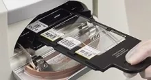
What is Whole Slide Imaging?
Whole Slide Imaging involves digitally scanning a tissue slide containing thin sections of tissue specimens for microscopic examination and storing it as digital images. This process allows for remote collaboration and analysis that can care about Image quality, ease of use, and speed.
How Does Whole Slide Imaging Work?
The process begins with the selection of an appropriate scanner and microscope. Once these have been chosen, slides are prepared according to standard protocols. The slides are then placed on the scanner’s stage and scanned using a motorized platform that moves across the surface at varying speeds depending on the resolution required. The image data accumulated during scanning is then processed using unique algorithms to create a digital version of the specimen that can be stored in a database. This image data can be shared with colleagues for collaboration and analysis.


