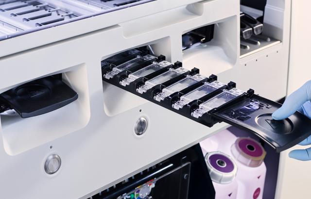
A Guide to Better IHC
This guide helps you identify your staining issue and provides potential remedies to assist you in troubleshooting the staining issue quickly.
Please keep in mind that some issues may be due to pre-staining conditions. Please refer to our 101 Steps to Better Histology guide for troubleshooting these pre-staining conditions.
In this guide, you will learn how to identify and troubleshoot some of the following common issues with IHC staining:
- No antibody staining is present
- No staining and circular artifact
- Loss of staining after staining is completed
- Staining too weak
- Background staining on glass slides
- Staining too strong/appears overstained
- Background staining or non-specific staining
- Uneven or gradient staining
- Atypical chromogen/counterstain staining
- Tissue artifacts
Additionally, receive 10 steps to optimizing your IHC staining.
Related Content
라이카 바이오시스템즈 Knowledge Pathway 콘텐츠는 에서 이용할 수 있는 라이카 바이오시스템즈 웹사이트 이용 약관의 적용을 받습니다. 법적고지. 라이카 바이오시스템즈 웨비나, 교육 프레젠테이션 및 관련 자료는 특별 주제 관련 일반 정보를 제공하지만 의료, 규정 또는 법률 상담으로 제공되지 않으며 해석되어서는 안 됩니다. 관점과 의견은 발표자/저자의 개인 관점과 의견이며 라이카 바이오시스템즈, 그 직원 또는 대행사의 관점이나 의견을 나타내거나 반영하지 않습니다. 제3자 자원 또는 콘텐츠에 대한 액세스를 제공하는 콘텐츠에 포함된 모든 링크는 오직 편의를 위해 제공됩니다.
모든 제품 사용에 다양한 제품 및 장치의 제품 정보 가이드, 부속 문서 및 작동 설명서를 참조해야 합니다.
Copyright © 2025 Leica Biosystems division of Leica Microsystems, Inc. and its Leica Biosystems affiliates. All rights reserved. LEICA and the Leica Logo are registered trademarks of Leica Microsystems IR GmbH.


