
Tips & Tricks to Multiplexing: How to Choose Chromogen Colors for Multiplex and Detection Systems for Multiplex Assays

Multiplexing is an important tool for simultaneous detection of multiple markers within a single tissue section.
Watch our 2 short videos below to learn more about multiplexing and download the complementary infographics to keep the points on hand when you need to revisit the information!
How to choose chromogen colors for multiplex
If you have opted for chromogenic multiplex staining, there are additional points to consider: the choice of colors and the order of color application. While this video won’t tell you exactly what, when, and where to use different chromogens, it will provide you with some tips to consider.
Check out below and learn with Larry from Leica Biosystems about the tips to consider for chromogenic multiplex staining into your tissue-based research.
10 Tips for Chromogenic Multiplexing
Chromogenic multiplex staining needs careful planning and consideration to produce a multi-color, multi-target, permanent slide that is resistant to photo-bleaching, and perfect for brightfield imaging and downstream analysis.
Consider using lighter chromogen colors
- Lighter chromogen colors may be easier on the eyes to visualize especially when multiple chromogens are involved.
Optimize the chromogen sequence
- DAB can overstain and occlude previously stained sites. Consider its suitable place in the assay.
Consider your choice of preferred counterstain
- Ensure the counterstain provides appropriate contrast and does not interfere with chromogen interpretation.
Consider the compatibility of your chromogens
- Consider the compatibility of your chosen chromogens with your required mounting media and preferred dehydration methods.
Consider the order of your markers
- Determine which antigens are robust or susceptible to degradation following multiple rounds of antigen retrieval
Test the stability of different chromogens
- Use chromogens with signals that remain strong earlier in the assay, and less robust signals later in the experiment.
Consider selecting the chromogen colors
- Consider selecting the chromogen colors for spatially close targets first and then build the assay further.
Consider the rate of chromogen precipitation
- Faster precipitating chromogens may be better suited for low expressed proteins. Consider slower precipitating chromogens for highly expressed proteins.
Consider high vs low expression
- Consider using a strong color chromogen for low expression, or low in quantity markers. Followed with a weaker color chromogen for high expression, or high in quantity markers.
Consider color combinations
- For colocalized markers, aim to use chromogens that will create a third color when combined.
Detection systems for multiplex assays
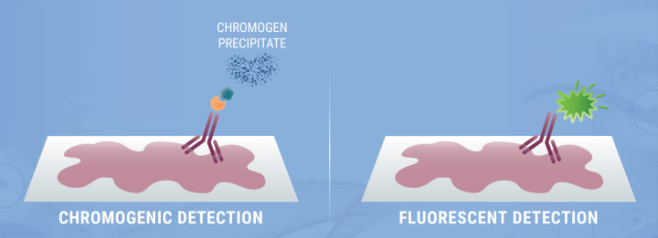
There are many decisions that must be made when adopting multiplex staining, such as choosing chromogenic or immunofluorescent multiplex assays, as well as the order and colors of chromogens and fluorophores. However, the decisions don’t stop here as there are different detection systems available for multiplex assays that can increase sensitivity and enhance the signal-to-noise ratio when detecting markers of interest. We will cover a few of these examples in this video.
Unsure of what detection systems are available for multiplex assays? Check out the infographic below that showcases some examples of detection systems available for multiplex assays that can increase sensitivity and enhance the signal-to-noise ratio when detecting markers of interest.
Multiplex Detection Systems
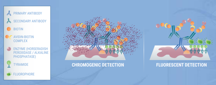
There are several detection systems available to visualize multiplex staining that are: direct primary antibody detection, or indirect detection with ABC, polymer-linked antibodies, with, or without tyramide amplification, and finally, oligonucleotide labelled primary antibodies with complementary probes. Whichever detection system you decide to use in your multiplex assay, optimization is vital.
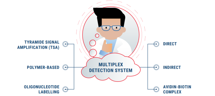
Direct Detection
- Suitable for highly expressed proteins
- Can avoid cross-reactivity by species-specific secondary antibodies
Indirect Detection
Tyramide Signal Amplification Detection System
Don't Miss Out! Subscribe To Research Link Today To Get Quaterly Updates On Our Latest Life Science Endeavours
For research use only. Not for use in diagnostic procedures.

발표자 소개
Rhian is a Scientist from Swansea University in Medical and Healthcare Studies and was featured in several collaborative publications. Rhian’s research-based background focused on tissue-based pathology in Multiple Sclerosis, primarily using immunohistochemical analysis and in vitro molecular techniques. She spent a short period conducting routine PCR testing for COVID-19 at the end of 2020.
Related Content


Leica Biosystems 콘텐츠는 Leica Biosystems 웹사이트 이용 약관의 적용을 받으며, 이용 약관은 다음에서 확인할 수 있습니다. 법적고지. 라이카 바이오시스템즈 웨비나, 교육 프레젠테이션 및 관련 자료는 특별 주제 관련 일반 정보를 제공하지만 의료, 규정 또는 법률 상담으로 제공되지 않으며 해석되어서는 안 됩니다. 관점과 의견은 발표자/저자의 개인 관점과 의견이며 라이카 바이오시스템즈, 그 직원 또는 대행사의 관점이나 의견을 나타내거나 반영하지 않습니다. 제3자 자원 또는 콘텐츠에 대한 액세스를 제공하는 콘텐츠에 포함된 모든 링크는 오직 편의를 위해 제공됩니다.
모든 제품 사용에 다양한 제품 및 장치의 제품 정보 가이드, 부속 문서 및 작동 설명서를 참조해야 합니다.
Copyright © 2025 Leica Biosystems division of Leica Microsystems, Inc. and its Leica Biosystems affiliates. All rights reserved. LEICA and the Leica Logo are registered trademarks of Leica Microsystems IR GmbH.