Fissazione e fissativi - Soluzioni di fissazione più diffuse

Fixation and Fixatives - Popular Fixative Solutions
In this fourth part of the Fixation and Fixatives series, we look at some of the many popular and traditional fixative solutions that have been used in histology for the last 100 years. This part also has an overview of proprietary solutions and provides advice on how to select the right fixative for your application.
Popular fixative solutions
Some of the more popular and traditional fixative solutions are shown below (click each line for details). This is by no means a complete list because during the past 100 years or more, there have been hundreds of variations to fixatives and fixative mixtures published. Those chosen are simply representative of the major groups. Some of these reagents can be purchased ready-to-use, from commercial suppliers. 1-4

1. Phosphate buffered formalin
Formulation
- 40% formaldehyde: 100 ml
- Distilled water: 900 ml
- Sodium dihydrogen phosphate monohydrate: 4 g
- Disodium hydrogen phosphate anhydrous 6.5 g
- The solution should have a pH of 6.8
- Fixation time: 12 – 24 hours
Recommended Applications
The most widely used formaldehyde-based fixative for routine histopathology. The buffer tends to prevent the formation of formalin pigment. Many epitopes require antigen retrieval for successful IHC following its use. Most pathologists feel comfortable interpreting the morphology produced with this type of fixative.
2. Formal calcium
Formulation
- 40% formaldehyde: 100 ml
- Calcium chloride: 10 g
- Distilled water: 900 ml
- Fixation time: 12 – 24 hours
Recommended Applications
Recommended for the preservation of lipids especially phospholipids.
3. Formal saline
Formulation
- 40% formaldehyde: 100 ml
- Sodium chloride: 9 g
- Distilled water: 900 ml
- Fixation time: 12 – 24 hours
Recommended Applications
This mixture of formaldehyde in isotonic saline was widely used for routine histopathology prior to the introduction of phosphate-buffered formalin. It often produces formalin pigment.
4. Zinc formalin (unbuffered)
Formulation
- Zinc sulfate: 1 g
- Deionized water: 900 ml
- Stir until dissolved then add –
- 40% formaldehyde: 100 ml
- Fixation time: 4 – 8 hours
Recommended Applications
Zinc formalin solutions were devised as alternatives to mercuric chloride formulations. They are said to give improved results with IHC. There are a number of alternative formulas available some of which contain zinc chloride, which is thought to be slightly more corrosive than zinc sulfate.
5. Zenker’s fixative
Formulation
- Distilled water: 950 ml
- Mercuric chloride: 50 g
- Potassium dichromate: 25 g
- Glacial acetic acid: 50 ml
- Fixation time: 4 – 24 hours
Recommended Applications
Gives good nuclear preservation but lyses red blood cells due to the presence of acetic acid. Has been recommended for congested specimens and gives good results with PTAH and trichrome staining. Produces mercury pigment which should be removed from sections prior to staining and can produce chrome pigment if tissue is not washed in water prior to processing. Is an intolerant agent, so, after water washing, tissue should be stored in 70% ethanol.
6. Helly’s fixative
Formulation
- Distilled water: 1000 ml
- Potassium dichromate: 25 g
- Sodium sulfate: 10 g
- Mercuric chloride: 50 g
- Immediately before use add –
- 40% formaldehyde: 50 ml
- Fixation time: 4 – 24 hours
Recommended Applications
Considered excellent for bone marrow, extramedullary hematopoiesis, and intercalated discs of cardiac muscle.
Produces mercury pigment which should be removed from sections prior to staining and can produce chrome pigment if tissue is not washed in water prior to processing. Is an intolerant agent, so, after water washing, tissue should be stored in 70% ethanol. Because of the low pH of this fixative formalin pigment may also occur.
7. B-5 fixative
Formulation
Stock solution
- Mercuric chloride: 12 g
- Sodium acetate anhydrous: 2.5 g
- Distilled water: 200 ml
Working solution, prepare immediately before use
- B-5 stock solution: 20 ml
- 40% formaldehyde: 2 ml
- Fixation time: 4 – 8 hours
Recommended Applications
Despite its mercury content and consequent problems with disposal, this fixative is popular for the fixation of hematopoietic and lymphoid tissue. It produces excellent nuclear detail, provides good results with many special stains, and is recommended for IHC. Sections will require the removal of mercury pigment prior to staining. Tissue should not be stored in this fixative but placed in 70% ethanol.
8. Bouin’s solution
Formulation
- Picric acid saturated aqueous solution. (2.1%): 750 ml
- 40% formaldehyde: 250 ml
- Acetic acid glacial: 50 ml
- Fixation time: 4 – 18 hours
Recommended Applications
Gives very good results with tissue that is subsequently trichrome stained. Preserves glycogen well but usually lyses erythrocytes. Sometimes recommended for gastrointestinal tract biopsies, animal embryos, and endocrine gland tissue. Stains tissue bright yellow due to picric acid. Excess picric should be washed from tissues prior to staining with 70% ethanol. Because of its acidic nature, it will slowly remove small calcium deposits and iron deposits.
9. Hollande’s
Formulation
- Copper acetate: 25 g
- Picric acid: 40 g
- 40% formaldehyde: 100 ml
- Acetic acid: 15 ml
- Distilled water: 1000 ml
Dissolve chemicals in distilled water without heat.
- Fixation time: 4 – 18 hours
Recommended Applications
Recommended for gastrointestinal tract specimens and fixation of endocrine tissues. Produces less lysis than Bouin. Has some decalcifying properties.
Fixative must be washed from tissues if they are to be put into phosphate-buffered formalin on the processing machine because an insoluble phosphate precipitate will form.
10. Gendre’s solution
Formulation
- 95% Ethanol saturated with picric acid: 800 ml
- 40% formaldehyde: 150 ml
- Acetic acid glacial: 50 ml
- Fixation time: 4 - 18 hours
Recommended Applications
This is an alcoholic Bouin solution that appears to improve upon aging. It is highly recommended for the preservation of glycogen and other carbohydrates. After fixation, the tissue is placed into 70% ethanol. Residual yellow color should be washed out before staining.
11. Clarke’s solution
Formulation
- Ethanol (absolute): 75 ml
- Acetic acid glacial: 25 ml
- Fixation time: 3 – 4 hours
Recommended Applications
Has been used on frozen sections and smears. Can produce fair results after conventional processing providing fixation time is kept very short. Preserves nucleic acids, but lipids are extracted. Tissues can be transferred directly into 95% ethanol.
12. Carnoy’s solution
Formulation
- Ethanol absolute: 60 ml
- Chloroform: 30 ml
- Acetic acid glacial: 10 ml
- Fixation time: 1 – 4 hours
Recommended Applications
Is rapid acting, gives good nuclear preservation, and retains glycogen. It lyses erythrocytes and dissolves lipids and can produce excessive hardening and shrinkage.
13. Methacarn
Formulation
- Methanol absolute: 60 ml
- Chloroform: 30 ml
- Acetic acid glacial: 10 ml
- Fixation time: 1 – 4 hours
Recommended Applications
Similar properties to Carnoy but causes less shrinkage and hardening.
14. Alcoholic formalin
Formulation
- 40% Formaldehyde: 100 ml
- 95% Ethanol: 900 ml
- 0.5 g calcium acetate can be added to ensure neutrality
- Fixation time: 12 - 24 hours
Recommended Applications
Combines a denaturing fixative with the additive and cross-linking effects of formalin. Is sometimes used during processing to complete fixation following incomplete primary formalin fixation. Can be used for fixation or post-fixation of large fatty specimens (particularly breast) because it will allow lymph nodes to be more easily detected as it clears and extracts lipids. If used for primary fixation, specimens can be placed directly into 95% ethanol for processing.
15. Formol acetic alcohol
Formulation
- Ethanol absolute: 85 ml
- 40% formaldehyde: 10 ml
- Acetic acid glacial: 5 ml
- Fixation time: 1 – 6 hours
Recommended Applications
A faster acting agent than alcoholic formalin due to the presence of acetic acid that can also produce formalin pigment. Sometimes used to fix diagnostic cryostat sections. If used for primary fixation, specimens can be placed directly into 95% ethanol for processing.
Proprietary fixative solutions
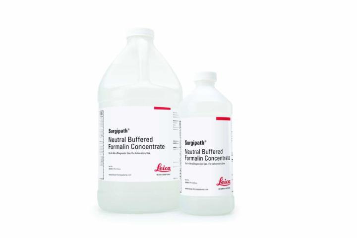
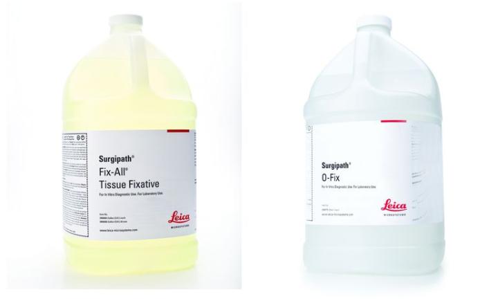
During the last few years, there has been an increasing number of proprietary fixatives developed for use in histopathology and medical research. They are generally marketed as less hazardous replacements for traditional formalin fixatives or as less toxic substitutes for fixative mixtures containing mercury such as B5.
Even though an MSDS must be provided, the exact composition of these reagents is not usually published and a potential user has to make do with a general description of the reagent. Those recommended as substitutes for B5 and Zenker’s (which are commonly used to fix lymphoid and hemopoietic tissues) usually contain zinc or barium salts and a low percentage of formaldehyde, while direct formalin substitutes often contain glyoxal and other components. It is in the latter group that reagents recommended for microwave-assisted fixation are found (see Part 5). Ethanol, methanol, and isopropanol are included in some formulations. 5

Which fixative should I use?
In most established laboratories, a routine fixative or fixatives have already been chosen and used for considerable time on a range of specimen types. The pathologist, histologist, or researcher will be completely accustomed to interpreting the characteristic tissue morphology produced by a particular fixative and processing schedule. Most frequently, the routine fixative will be neutral buffered formalin with other agents used for bone marrow trephines (perhaps a zinc formalin), renal biopsies, frozen sections, etc. Buffered formalin is widely used because it is probably the most flexible of agents. It can be incorporated into the processing schedule on enclosed tissue processors. It permits the successful application of a wide range of special stains. Immunohistochemistry methods, which generally include an antigen retrieval step, have been optimized for formalin-fixed tissues, and tissue specimens can be stored in formalin for extended periods without major deleterious effects. Molecular techniques such as ISH have also been validated for use on formalin-fixed tissue. It is against these characteristics that a new or replacement fixative must be judged.5-6
Some laboratories are currently looking to replace formalin with a less toxic reagent, and there are several alternatives described in the previous paragraphs. In a research environment, where a specific tissue element is being studied, there may be more control over the fixation step, and it is worthwhile testing several reagents before making a final decision. If you are contemplating a change of fixative, consider the following properties in addition to evaluating the results you see under the microscope:
- Toxicity of the fixative (both short-term and cumulative)
- Volatility of its components and the equipment available to prevent staff exposure to the agent
- Flammability
- The effect of over-fixation on tissues (does prolonged fixation damage tissues?)
- Storage requirements if specimens cannot be left in fixative
- The compatibility of the fixative with your tissue processor (might it damage components?)
- The practical and legal requirements of disposal after use
Changing to a different fixative requires very careful consideration and thorough evaluation.
About the presenter

Geoffrey Rolls is a Histology Consultant with decades of experience in the field. He is a former Senior Lecturer in histopathology in the Department of Laboratory Medicine, RMIT University in Melbourne, Australia.
References
- Eltoum I, Fredenburgh J, Myers RB, Grizzle WE. Introduction to the theory and practice of fixation of tissues. J Histotechnol 2001;24;173 -190.
- Leong AS-Y. Fixation and fixatives. In Woods AE and Ellis RC eds. Laboratory histopathology. New York: Churchill Livingstone, 1994;4.1-1 - 4.1-26.
- Hopwood D. Fixation and fixatives. In Bancroft J and Stevens A eds. Theory and practice of histological techniques. New York: Churchill Livingstone, 1996.
- Carson FL. Histotechnology. 2nd ed. Chicago: ASCP Press, 2007.
- Titford ME, Horenstein MG. Histomorphologic Assessment of Formalin Substitute Fixatives for Diagnostic Surgical Pathology. Arch Pathol Lab Med 2005;129;502-506.
- Kothmaier H, Rohrer D, Stacher E, Quehenberger F, Becker K-F, Popper HH. Comparison of Formalin-free Tissue Fixatives: A Proteomic Study Testing Their Application for Routine Pathology and Research. Arch Pathol Lab Med 2011;135;744-752.
Related Content
Leica Biosystems Knowledge Pathway content is subject to the Leica Biosystems website terms of use, available at: Legal Notice. The content, including webinars, training presentations and related materials is intended to provide general information regarding particular subjects of interest to health care professionals and is not intended to be, and should not be construed as, medical, regulatory or legal advice. The views and opinions expressed in any third-party content reflect the personal views and opinions of the speaker(s)/author(s) and do not necessarily represent or reflect the views or opinions of Leica Biosystems, its employees or agents. Any links contained in the content which provides access to third party resources or content is provided for convenience only.
For the use of any product, the applicable product documentation, including information guides, inserts and operation manuals should be consulted.
Copyright © 2025 Leica Biosystems division of Leica Microsystems, Inc. and its Leica Biosystems affiliates. All rights reserved. LEICA and the Leica Logo are registered trademarks of Leica Microsystems IR GmbH.


