Science of H&E

Download this training resource to learn more about routine staining with hematoxylin and eosin (H&E) and the steps involved with the staining process.
Why Do We Stain?
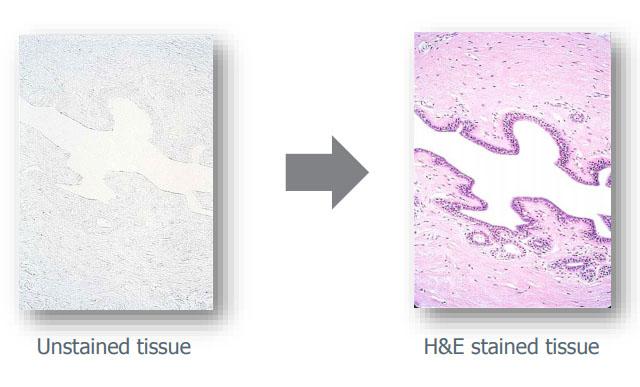
In order to deliver a medical diagnosis, tissues must be examined under a microscope. Once a tissue specimen has been processed by a histology lab and transferred onto a glass slide, it needs to be appropriately stained for microscopic evaluation. This is because unstained tissue lacks contrast: when viewed under the microscope, everything appears in uniform dull grey color.

What Does "Staining" Do?
- Contrasts different cells
- Highlights particular features of interest
- Illustrates different cell structures
- Detects infiltrations or deposits in the tissue
- Detects pathogens

About the presenter

Andrew Lisowski has almost 30 years of experience in histology and histotechnology. He attended veterinary school and earned his master’s degree in molecular biology. Andrew worked in histology, IHC and ISH labs, cell culture lab, performed in-vitro and in-vivo toxicology assays and was a member of a necropsy team. He worked for pharmaceutical companies, medical school and founded his own molecular and histology firms.
Related Content
El contenido de Leica Biosystems Knowledge Pathway está sujeto a las condiciones de uso del sitio web de Leica Biosystems, disponibles en: Aviso legal.. El contenido, incluidos los webinars o seminarios web, los recursos de formación y los materiales relacionados, está destinado a proporcionar información general sobre temas concretos de interés para los profesionales de la salud y no está destinado a ser, ni debe interpretarse como asesoramiento médico, normativo o jurídico. Los puntos de vista y opiniones expresados en cualquier contenido de terceros reflejan los puntos de vista y opiniones personales de los ponentes/autores y no representan ni reflejan necesariamente los puntos de vista ni opiniones de Leica Biosystems, sus empleados o sus agentes. Cualquier enlace incluido en el contenido que proporcione acceso a recursos o contenido de terceros se proporciona únicamente por comodidad.
Para el uso de cualquier producto, debe consultarse la documentación correspondiente del producto, incluidas las guías de información, los prospectos y los manuales de funcionamiento.
Copyright © 2025 Leica Biosystems division of Leica Microsystems, Inc. and its Leica Biosystems affiliates. All rights reserved. LEICA and the Leica Logo are registered trademarks of Leica Microsystems IR GmbH.


