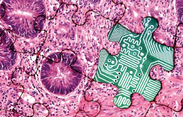
Creating Digital Ready Slides - A Practical Guide

The adoption of digital pathology is a multifaceted project involving many stakeholders across the pathology department. The impact on the laboratory is not isolated to simply installing a scanner but rather affects the whole workflow to generate optimized Digital Ready Slides. Standardization of histological slide preparation requires focusing on optimizing individual workflow steps and a holistic overview of the complete process from sample acquisition right through to diagnosis. Knowing this in advance and taking appropriate steps to support effectively change management can promote engagement and pave a path to success.
Key Learning Objectives:
- Define the critical attributes of Digital Ready Slides
- Demonstrate the impact of tissue preparation steps on scan quality
- Provide practical guidance to creating Digital Ready Slides and additional considerations at each laboratory step
About the presenter

Dr. Colgan has over a decade of experience in the digital pathology sector and is focused on how this new and disruptive technology can be leveraged to provide real benefits in both the healthcare and research domains. Prior to working with Leica Biosystems, she came from a research background with a BSc in Biotechnology and a PhD in Vascular Biology from Dublin City University, Ireland.
Referencias
- Fraggetta, Filippo & Garozzo, Salvatore & Zannoni, GianFranco & Pantanowitz, Liron & Rossi, EstherDiana. (2017). Routine Digital Pathology Workflow: The Catania Experience. Journal of Pathology Informatics. 8. 51. 10.4103/jpi.jpi_58_17.
- Williams, Bethany & Hanby, Andrew & Millican‐Slater, Rebecca & Nijhawan, Anju & Verghese, Eldo & Treanor, Darren. (2017). Digital Pathology for the Primary Diagnosis of Breast Histopathological Specimens: An Innovative Validation and Concordance Study. Histopathology. 72. 10.1111/his.13403.
- Hanna, Matthew & Reuter, Victor & Ardon, Orly & Kim, David & Sirintrapun, Sahussapont & Schüffler, Peter & Busam, Klaus & Sauter, Jennifer & Brogi, Edi & Tan, Lee & Xu, Bin & Bale, Tejus & Agaram, Narasimhan & Tang, Laura & Ellenson, Lora & Philip, John & Corsale, Lorraine & Stamelos, Evangelos & Friedlander, Maria & Hameed, Meera. (2020). Validation of a digital pathology system including remote review during the COVID-19 pandemic. Modern Pathology. 33. 10.1038/s41379-020-0601-5.
- College of American Pathologists, “COVID-19 – Remote Sign-Out Guidance” (2020). Available at: capatholo.gy/3ghf4Wu.
- Centers for Medicare & Medicaid Services, “Clinical Laboratory Improvement Amendments (CLIA) Laboratory Guidance During COVID-19 Public Health Emergency” (2020). Available at: go.cms.gov/2LGD2wh.
- Royal College of Pathologists, “Guidance for remote reporting of digitalpathology slides during periods of exceptional service pressure” (2020). Available at: https://www.rcpath.org/profession/digital-pathology.html
- McCarthy, J. & Minsky, M.L. & Rochester, N. & Shannon, C.E.. (1955).proposal for the Dartmouth summer research project on artificial intelligence. Artificial Intelligence: Critical Concepts. 2. 44-53.
- Kanavati, Fahdi & Toyokawa, Gouji & Momosaki, Seiya & Rambeau, Michael & Kozuma, Yuka & Shoji, Fumihiro & Yamazaki, Koji & Takeo, Sadanori & Iizuka, Osamu & Tsuneki, Masayuki. (2020). Weakly-supervised learning for lung carcinoma classification using deep learning. Scientific Reports. 10.10.1038/s41598-020-66333-x.
- Iizuka, Osamu & Kanavati, Fahdi & Kato, Kei & Rambeau, Michael & Arihiro, Koji & Tsuneki, Masayuki. (2020). Deep Learning Models for Histopathological Classification of Gastric and Colonic Epithelial Tumours. Scientific Reports. 10. 10.1038/s41598-020-58467-9.
- McCampbell, Adrienne & Raghunathan, Varun & Tom-Moy, May & Workman, Richard & Haven, Rick & Ben-Dor, Amir & Rasmussen, Ole & Jacobsen, Lars & Lindberg, Martin & Yamada, N. & Schembri, Carol. (2017). Tissue Thickness Effects on Immunohistochemical Staining Intensity of Markers of Cancer. Applied Immunohistochemistry & Molecular Morphology. 27. 1. 10.1097/PAI.0000000000000593.
- Masuda, Shinobu & Suzuki, Ryohei & Kitano, Yuriko & Nishimaki, Haruna & Kobayashi, Hiroko & Nakanishi, Yoko & Yokoi, Hideo. (2020). Tissue Thickness Interferes With the Estimation of the Immunohistochemical Intensity: Introduction of a Control System for Managing Tissue Thickness. Applied Immunohistochemistry & Molecular Morphology. Publish Ahead of Print. 1. 10.1097/PAI.0000000000000859.
- Arif, Ahmed & Stuerzlinger, Wolfgang. (2009). Analysis of text entry performance metrics. TIC-STH’09: 2009 IEEE Toronto International Conference - Science and Technology for Humanity. 100 - 105. 10.1109/TICSTH.2009.5444533.
Related Content
El contenido de Leica Biosystems Knowledge Pathway está sujeto a las condiciones de uso del sitio web de Leica Biosystems, disponibles en: Aviso legal.. El contenido, incluidos los webinars o seminarios web, los recursos de formación y los materiales relacionados, está destinado a proporcionar información general sobre temas concretos de interés para los profesionales de la salud y no está destinado a ser, ni debe interpretarse como asesoramiento médico, normativo o jurídico. Los puntos de vista y opiniones expresados en cualquier contenido de terceros reflejan los puntos de vista y opiniones personales de los ponentes/autores y no representan ni reflejan necesariamente los puntos de vista ni opiniones de Leica Biosystems, sus empleados o sus agentes. Cualquier enlace incluido en el contenido que proporcione acceso a recursos o contenido de terceros se proporciona únicamente por comodidad.
Para el uso de cualquier producto, debe consultarse la documentación correspondiente del producto, incluidas las guías de información, los prospectos y los manuales de funcionamiento.
Copyright © 2025 Leica Biosystems division of Leica Microsystems, Inc. and its Leica Biosystems affiliates. All rights reserved. LEICA and the Leica Logo are registered trademarks of Leica Microsystems IR GmbH.


